
Wednesday, August 12, 2015

Friday, June 26, 2015
Wednesday, May 22, 2013
Malignant Colon Cancer
Adenocarcinoma of the colon
Risk factors
- Low-fiber diet
- Diet low in fruits and vegetables
- High red meat and animal fat consumption
- Adenomatous polyps
- Hereditary polyposis syndromes
- Lynch syndrome
- Ulcerative colitis
Genetics
- Multiple mutations are involved
- APC gene
- K-ras oncogene
- p53 gene
Pathology
Microscopically:
It is columnar cell carcinoma originating in the colonic epithelium.
Macroscopically: 4 forms
Annular- may give rise to obstructive symptoms
Tubular- presents with bleeding
Ulcer-presents with bleeding
Cauliflower- least malignant-presents with bleeding
Tumors are more common in the left colon and rectum.
Spread
Direct Spread:
Locally it can invade the bladder, obstruct ureter and may cause hydronephrosis.
Can perforate and cause peritonitis/pericolic abscess/ faecal fistula.
Lymphatic spread:
Growth through lymphatics spreads to pericolic, epicolic, intermediate and principal group of lymph nodes.
Bloodstream spread: To the liver via portal system
Staging colon cancer: Modified Dukes' Staging
Stage
A
B1
B2
C1
C2
D
Extent of Disease
Limited to the mucosa and submucosa
Invasion into but not through the muscularis propria
Invasion through the muscularis propria
Positive lymph nodes; invasion into but not through the muscularis propria
Positive lymph nodes; invasion through the muscularis propria
Distant metastasis
Clinical features
Age: common in patients over 50 yrs of age.
Carcinoma of the left side of the colon:
Most tumors occur in this site and are of stenosing variety.
Main sympton- increasing intestinal obstruction.
Pain
Pain is referred to suprapubic area. Pt. have episodic colicky pain. Constant pain suggests advanced tumors.
Altered bowel habit:
The episodes of constipation may be followed by attacks of diarrhoea.
Palpable lump
Distension: Lower abdominal distension.
- Carcinoma of sigmoid
Same features as above with there variations:
Pain
Pain
Tenesmus
Low tumors give rise to a feeling of the need for evacuation, which result in tenesmus accompanied by the passage of mucus (esp. in the early morning)
Bladder symptoms
Carcinoma of transverse colon:
May be mistaken for a carcinoma of the stomach because of the position of the tumor together with anemia and lassitude.
Carcinoma of the caecum and ascending colon:
Anemia
Presence of mass in the right iliac fossa.
Can present with obstruction
Metastatic disease:
Pt. may also present for the first time with liver metastases and an enlarged liver, ascites,
Rarely pt. may also present metastases to the lung, skin, bone and brain.
Right V/S left sided cancer
Right-Sided Cancer
Gross
Polypoid mass
Barium studies
Polypoid mass
Presentation
Bleeding
Occult blood in stool
Iron deficiency anemia
Left-Sided Cancer
Gross
Circumferential growth producing a "napkinring“ Configuration
Barium studies
"Apple core" lesion
Presentation
Change in bowel habits
Constipation or diarrhea
Reduced caliber stools
Obstruction
Investigations
Sigmoidoscopy
It is a part of the routine investigation of patients passing blood and mucus if it limited to rectum.
Colonoscopy
Used in pts. With presenting symptom of bleeding, those with polyps and in those with radiological doubt.
Radiology
Double-contrast barium enema is used
It shows irregular filling defect and apple core lesion( in lt. sided Ca)
Ix of choice in pt. with a predominant change in bowel habit as a presenting symptom.
USG
For screening of liver metastases
CT
to determine the local invasion in case of large palpable abdominal mass.


Treatment
For right sided growth
Right radical hemicolectomy with ileo transverse anastomosis is done.
Structures removed are-terminal 30cm of ileum, caecum, appendix, ascending colon, 1/3 transverse colon and lymph nodes (epicolic, paracolic, intermediate)
In inoperable right sided growth, ileo-transverse anastomosis is done as a bypass procedure.
For left sided growth
Left radical hemicolectomy is done.
Structures removed are- left ½ of tansverse colon and descending colon is removed along with lymph nodes.
Left sided stenosing type of growth can present with acute intestinal obstruction, in which case initially colostomy is done. Later after 3-6 wks, following proper preparation, required procedure is done, followed by closure of colostomy after 8 wks.
In solitary liver secondary
Segmental hepatic resection is done.
In case of multiple secondaries confined to one lobe of the liver, hemihepatectomy can be done.
Adjuvant therapy
Chemotherapy using 5-FU and cyclophosphamide.
Regimes-Levamisole and 5-FU or Leucoverin and 5-FU
Indications-Positive nodes, T4 lesion, Venous spread, Signet cell type, Poorly differentiated, Changes in CEA level.
Hepatic metastases
Hepatic resection after recovery from colonicresection.
Irresectable symptomatic hepatic metastases may be treated with cytotoxic drugs or ablaative treatments.
.png)
.png)
.png)

Monday, May 20, 2013
Thyroid Neoplasms
“CLASSIFICATION”
Benign: follicular adenoma
Malignant:
1. Primary-
2. Secondary
Follicular Ca. surgery
Minimal capsular invasion, <1cm---lobectomy
Vascular invasion---Total thyroidectomy
Anaplastic Ca.
Surgery rarely indicated.
L/N surgery
Neck surgery
Malignant:
1. Primary-
a. Follicular epithelium ( differentiated)
Follicular
Papillary
b. Follicular epithelium (undifferentiated)
Anaplastic
c. Parafollicular cells
Medullary
d. Lymphoid cells
Lymphoma
2. Secondary
Metastatic
Papillary Carcinoma
a. Epidemiology
Accounts for 80% of malignant thyroid tumors.
F>M ; Age 20-50
Risk factor: radiaton exposure
b. Micro
exhibits a papillary pattern.
psammoma bodies present.
characteristic nuclear features:
clear “Orphan Annie eye” nuclei.
nuclear grooves.
intranuclear cytoplasmic inclusions.
c. Lymphatic spread to cervical nodes is commom.
d. Treatment: Resection is curative in most cases.
Radiotherapy with iodine 131 is effective for metastases.
e. Prognosis: excellent
Follicular carcinoma
a. Accounts for 15% of malignant thyroid tumors
b. Females> males; age 40-60
c. Hematogenous metastasis to the bones or lungs is common
Medullary carcinoma
a. Accounts for 5% of malignant thyroid tumors
b. Arises from C cells (parafollicular cells) and secretes calcitonin
c. Micro: nests of polygonal cells in an amyloid stroma
d. Minority (25%) are associated with MEN II and MEN III syndromes
Anaplastic carcinoma
a. Presentation
i. Females> males; age >60
ii. Firm, enlarging, bulky mass
iii. Dyspnea and dysphagia
iv. Tendency for early widespread metastasis and invasion of the trachea and esophagus
b. Micro: undifferentiated, anaplastic, and pleomorphic cells
c. Prognosis: very aggressiveand rapidly fatal
Surgical terms:
Lobectomy: complete removal of one lobe including isthmus.
Near total thyroidectomy: Total lobectomy with isthmusectomy with subtotal lobectomy.
Subtotal thyroidectomy: isthmusectomy with subtotal lobectomy b/l
Total thyroidectomyb/l lobectomy with isthmusectomy.
Management of thyroid cancers
Papillary Ca. thyroid surgery:
- Node negative, <1cm---lobectomy
- >1cm----Total thyroidectomy
- Evidence of spread present----Total thyroidectomy
- Familial disease---Total thyroidectomy
- Neck irradiation in childhood---Total thyroidectomy
Follicular Ca. surgery
Minimal capsular invasion, <1cm---lobectomy
Vascular invasion---Total thyroidectomy
Anaplastic Ca.
Surgery rarely indicated.
L/N surgery
Neck surgery
Tuesday, March 5, 2013
RENAL TUMORS
Classification


.png)

Treatment:
Renal Cell carcinoma


 Sarcomatoid


.png)
Benign
- Cortical adenoma
- Angiomyolipoma
Malignant
- RCC
- Upper tract TCC
- Nephroblastoma
- Squamous CA of renal pelvis
Cortical adenoma
- Discovered incidentally, mostly found on autopsy
- Less than 1 cm without malignant features (pea like)
- Symptoms are unusual
Microscopically uniform basophilic or acidophilic with monotonous nuclear and cellular composition

Renal angiomyolipoma
- May occur alone or a part of tuberous sclerosis
- Hamartomas composed of fat, smooth muscle, and blood vessels
- AML may be found in the eyes, heart, lung and bone


- Malignant Tumors
Wilms tumor (nephroblastoma)
Peak age:2-5 years
Risk factors
. WAGR syndrome-Wilms tumor, aniridia, genital anomalies, and mental retardation
. Beckwith-Wiedemann syndrome
.Tumor suppressor genes
. WT-l (llp13)
. WT-2 (llp15)
Gross: large solitary tan mass
.png)
Micro
1. Metanephric blastema
ii. Epithelial elements (immature glomeruli and tubules)
iii. Stroma

C/F:
Abdominal mass
Pyrexia
Hematuria
Metastasis occurs early, mainly to lungs.
Inv:
USG
Urography or
CT
Nephrectomy followed by radiation with or without chemotherapy
If B/L tumor: Partial Nephrectomy
Prognosis: excellent; long-term survival rate of 90%
Epidemiology
- Male: Female 2:1
- 40-60 years of age
Risk factors
i. Cigarette smoking
ii. Chronic analgesic use
iii. Asbestos exposure
iv. Chronic renal failure and acquired cystic disease
v. Von Hippel- Lindau disease
Gross
. Large solitary yellow mass found in the poles (most commonly in the upper pole: most common site- PCT)
. Areas of necrosis and hemorrhage are commonly present
. The tumor often invades the renal vein and may extend into the vena cava and heart


Pathology
Clear cell types
- more than 80%
- polygonal or round cells with clear cytoplasm

Papillary variant
- 14% of RCC
- macroscopic feature-small, confined to cortex and nearly completely encapsulated
- Micro: papillae covered by small cells with scanty cytoplasm, arranged in a single layer on the papillary basement membrane with low nuclear grade

- aggressive behavior and poor prognosis
- microscopic features- spindle cell pattern

Chromophobe variant
- 4% of RCC
- microscopic features light and transparent cytoplasm
- better prognosis

Signs and symptoms
Classic triad (pain, lump and hematuria) is found only in 10% cases
Weight loss, fever, hematuria, night sweats and sudden development of varicocele
Paraneoplastic syndromes from ectopic hormone production
. Polycythemia(erythropoietin production)
. Hypertension (renin production)
. Cushing syndrome (corticosteroid synthesis)
. Hypercalcemia (PTH-like hormone)
. Feminization or masculinization (gonadotropin release)
High incidence of metastasis on initial presentation
May cause amyloidosis, a leukemoid reaction, or eosinophilia
Investigations
IVU


CT scan

.png)
- Renal Angiogram
- CX-Ray
- Bone scan
Treatment
- RCC is highly resistant to radio and chemotherapy
- Nephrectomy with removal of the perinephric fat
LUNG TUMORS

Bronchogenic carcinoma
- Epidemiology
- Leading cause of cancer death among both men and women
- Increasing in women (increased smoking) in the past few decades
- Occurs most commonly from 50-80 years of age
i. Cigarette smoking
ii. Occupational exposure (asbestosis, uranium mining, radiation, etc.)
iii. Air pollution
Common genetic mutations
i. Oncogenes
L-myc: small cell carcinomas
K-ras: adenocarcinomas
ii. Tumor suppressor genes p53 and the retinoblastoma gene
- Cough
- sputum production
- weight loss
- anorexia
- fatigue
- dyspnea
- hemoptysis and
- chest pain
- hoarseness of voice
- Obstruction may produce focal emphysema, atelectasis, bronchiectasis or pneumonia
.Males=females
.less closely associated with smoking than squamous cell
.Gross: peripheral gray-white mass with pleural puckering

.May develop in areas of parenchymal scarring (scar carcinoma)
.Micro: tumor forms glands and may produce mucin.

Bronchioloalveolar carcinoma (5%)
. Subset of adenocarcinoma
. Arises from terminal bronchioles or alveolar walls
. Gross: peripheral mucinous gray-white nodules

Micro:
columnar tumor cells grow along the walls of pre-existing alveoli

Squamous cell carcinoma (30%)
. Males> females
. strongly related to smoking
Gross: usually centrally located, gray-white bronchial mass
Arises from bronchial epithelium after a progression:
metaplasia ~dysplasia ~ carcinoma in situ ~ invasive carcinoma

Micro:
. Invasive nests of squamous cells
. Intercellular bridges (desmosomes)
. Keratin production ("squamous pearls")

Small cell (oat cell) carcinoma (20%)
. Males> females
. strong association with smoking
Very aggressive: rapid growth and disseminate early
Gross: central, gray-white masses
+carcinoma.png)
Micro:
small round or polygonal cells in clusters

EM: cytoplasmic dense-core neurosecretory
granules

Commonly associated with paraneoplastic syndromes.
Also most common cause of venacaval obstruction syndromes.
Large cell carcinoma (10%)
In early stages, is associated with cavitation
Gross: peripherally located lesion

Micro:
large anaplastic cells without evidence of differentiation

Intrathoracic spread
i. Lymph nodes (50%):
hilar, bronchial, tracheal, and mediastinal
ii. Pleural involvement (adenocarcinoma)
iii. Pancoast tumor (apical tumor) causing Horner syndrome
iv. Superior vena cava syndrome
Obstruction of SVC by tumor
.Distended head and neck veins
.Plethora
.Facial and upper arm edema
v. Esophageal obstruction: dysphagia
vi. Recurrent laryngeal nerve involvement: hoarseness
vii. Phrenic nerve damage: diaphragmatic paralysis
Extrathoracic sites of metastasis:
adrenal (>50%)
liver
brain and
bone
Paraneoplastic syndromes
i. Endocrine/metabolic syndromes
. ACTH ~ Cushing syndrome
. ADH~ SIADH
. PTH ~ hypercalcemia (squamous cell carcinomas)
ii. Eaton-Lambert syndrome
iii. Acanthosis nigricans
iv. Hypertrophic pulmonary osteoarthropathy
. Periosteal new bone formation
. Clubbing
. Arthritis
Investigations
Sputum cytology
Bronchoscopy:
Best for centrally located lesions
Fine Needle Aspiration Bx
for peripheral lesions
Pleural Bx in all patients presenting with pleural effusion.
CX-Ray: common features-
a. U/L hilar lymphadenopathy
c. Lung, lobe or segmental collapse
d. Pleural effusion

b. Peripheral pulmonary opacity

c. Lung, lobe or segmental collapse

d. Pleural effusion

f. Rib destruction
CT Chest
Others:
CT head
Liver ultrasound
Bone marrow biopsy
Treatment
1.Surgical resection
Symptoms that suggest unresectable lesion: Wt. loss >10%
- Bone pain
CNS symptoms
Tumor involving trachea, esophagus, pericardium and chest wall.
Small cell CA
2. Radiotherapy:
palliation of distressing complications like SVC obstruction, rec. hemoptysis, pain caused by chest wall invasion.
Used in adjunct to Chemothrapy for small cell ca.
3. Chemotherapy
esp. used in small cell CA along with RT.
I.v Cyclophophamide, doxorubicin and vincristine
OR
I.v Etoposide and Cisplatin are used.
4. Neoadjuvant and adjuvant CT
CT given surgery for down staging the disease in non small cell CA.
Post-op CT used if lymph node is involved
5. Effusion sclerosed with tetracycline.
Prognosis:
Poorest for small cell carcinoma
Best after surgical resection of squamous cell CA.
Blood borne metastatic deposits from many primary tumors: part. Breast
kidney
uterus
ovary
testes
thyroid
Deposits are usually multiple and bilateral.
Often there are no respiratory symptoms and diagnosis is made by radiological examination.

Malignant mesotheliomas
. Rare highly malignant neoplasm affecting the pleura.
. Occupation exposure to asbestos in 90% of cases
. Presents with recurrent pleural effusions, dyspnea, chest pain
. Gross: encases and compresses the lung
. Micro: carcinomatous and sarcomatous elements (biphasic pattern)
. EM: long, thin microvilli
.No curative treatment and chain pain is often difficult to control.
. Poor prognosis.
Saturday, June 16, 2012
CNS TUMORS
Epidemiology
Half of all brain and spinal cord tumors are metastatic
Most frequent primary CNS tumors: Meningiomas
Glioblastoma multiforme
Clinical manifestations
Headache, often worse at night or early morning
Seizures, with tumors involving cerebral cortex
Mental changes (e.g., deficits in memory, concentration, reasoning, etc.)
Focal neurological symptoms, related to involvement of specific brain regions
Symptoms related to increased intracranial pressure
i. Presence of a space-occupying mass within the cranial cavity
ii. Blockage of CSF flow
iii. Edema around the tumor (peri tumoral edema)
Special features of brain tumors
i. Malignant CNS tumors do not metastasize outside the cranial cavity.
ii. Clinical consequences depend on infiltrative behavior and location.
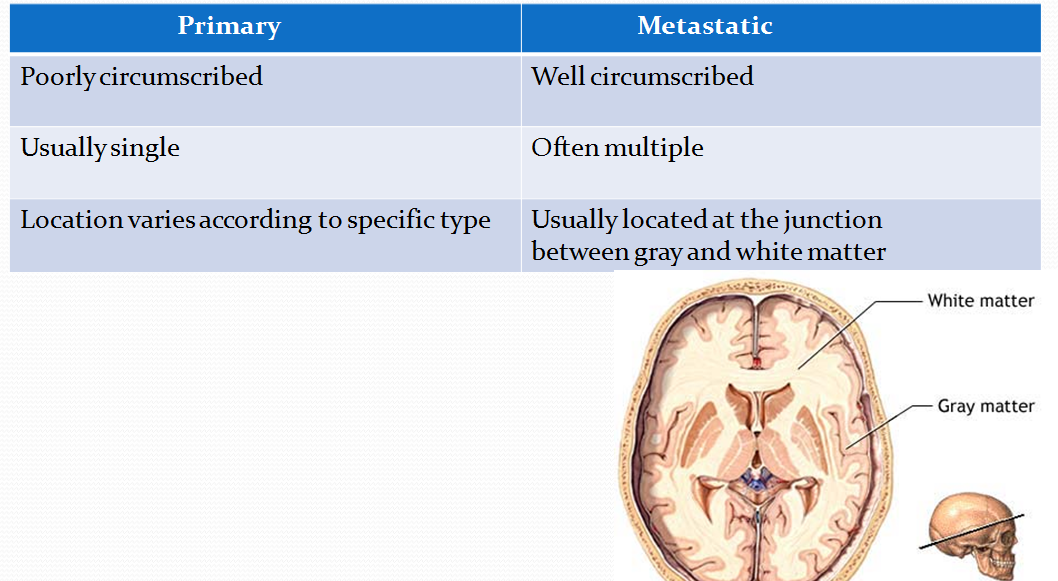
Differences Between Primary and Metastatic Tumors
Astrocytomas
a. Originate from astrocytes and exhibit
i. Fibrillary background
ii. Immunoreactivity for glialfibrillary acidic protein (GFAP)
iii. Diffuse (ill-demarcated) pattern of growth
a. Fibrillary astrocytomas
Grading is important for both prognosis and treatment. Most frequent systems
Four grades based on nuclear atypia (pleomorphism),
-mitoses, necrosis, and vascular endothelial hyperplasia (VEH)
Grade 1-2 astrocytomas are well differentiated astrocytomas
Grade 3 astrocytomas are anaplastic astrocytomas
Grade 4 astrocytomas are called glioblastoma multiforme (GBM)
GBM is the most cormmon CNS primary malignancy
Most common location: white matter
Histology: marked nuclear atypia, mitoses, necrosis, and VEH
Characteristic histo pathological feature: pseudopalisading necrosis
VEH is often florid, giving rise to glomeruloid formations

Glioblastoma multiforme has a tendency to cross the midline
by involving the corpus callosum, hence k/a"Butterfly glioma"

Anaplastic astrocytoma showing marked nuclear pleomorphism

b. Pilocytic astrocytoma
Benign astrocytic tumor of children and young adults
Locations: posterior fossa (cerebellum) and diencephalon
Often presents as a cystic lesion with a mural nodule


Histology:
-spindly neoplastic astrocytes with long bipolar processes
-tumors rich in Rosenthal fibers (thick corkscrew-like eosinophilic structures, which derive from hypertrophic processes of astrocytes)
Favorable prognosis for posterior fossa tumors

Oligodendroglioma
Glioma of oligodendroglial origin
Occurs in 30- to 50-year-old patients
Location: white matter of cerebral hemispheres adjacent to neocortex

Often manifests with seizures
Characteristic histopathology
i. Neoplastic cells are similar to oligodendroglia
ii. Pronounced perinuclear halo: "fried-egg" appearance

iii. Prominent capillary network in a chickenwire pattern
Slow-growing tumors that allow long survival (average 5-10 years)
Recur after surgery and degenerate into high-grade gliomas over time

Ependymoma
Glioma of ependymal origin
Location
1. Children: fourth ventricle
ii. Adults: lateral ventricle or spinal canal
Gross appearance: circumscribed tumors with papillary architecture

Histology: neoplastic cells resemble ependymal cells. Characteristic features:
i. Ependymal rosettes

ii. Perivascular pseudo rosettes

Often presents with obstructive hydrocephalus, when present in the fourth ventricle
Tend to recur after surgery and acquire more aggressive behavior
Meningioma
Originates from meningothelial cells of the arachnoid
Tumors of adulthood (women> men), rare in children
May develop at any meningeal site. Most frequent are dural convexities
Gross: attached to the dura,
pushes underlying brain
without invasion

Microscopic
i.Spindle-shaped cells with indistinct borders (syncytial)
ii. Cells arranged in whorls or fascicles
iii. Psammoma bodies frequent


Generally, good prognosis
Tumors in some location may not be amenable to complete resection
Primitive neuroectodermal tumors (PNET)
Highly undifferentiated; originate from a primordial neuroglial precursor
Variably named, depending on location in the brain
Most frequent PNETs: medulloblastoma and
retinoblastoma
All PNETs share the following features:
i. Develop in children
ii. Histology: blue, small, round cell tumors, with pseudorosettes
iii. Highly aggressive but responsive to radiation therapy
.
Histo:

Medulloblastoma arises in the cerebellar vermis (midline location)
i. Grows rapidly and spreads through CSF
ii. Resection and radiation therapy allow 5-year survival of 75%.

Schwannoma
Originates from Schwann cells of cranial or spinal nerves
Associated with NF type II
Most frequent location: eighth cranial nerve
cerebellopontine angle(CPA)
Manifests characteristically with loss of hearing and tinnitus

Histology
i. Spindle cells arranged in hypercellular Antoni A areas, alternating with hypocellular Antoni B areas
ii. Verocay bodies: parallel rows of neoplastic Schwann cells

Verocay bodies
Neoplastic cells are immunoreactive for a protein called S-100
Good prognosis after surgical resection

Craniopharyngioma
Arises from the remmanants of Rathke’s pouch
Patients affected are usually children or young adults
Location: within the suprasellar/diencephalic region
Gross: craniopharyngiomas are cystic or partially-cystic with solid areas.

Histo: nesting of squamous epithelium bordered by radially arranged cells.
resembles adamantinoma, the most common tumor of the tooth

Benign but tends to recur after resection
T/T:
Trans-sphenoidal resection of the tumor.
Investigations of Brain tumors:
1. CT/MRI Head
allows localization of the tumor.
MRI has value in investigation of posterior fossa tumors and brain stem
2. CXR:
provides evidence of primary lung tumor
T/t
1.Medical: Relief of ICP is reqd. when surgery is not possible or
when life is threatened before diagnosis is revealed.
Dexamethasone is used.
2. Surgical:
Main stay of t/t
3. Radiotherapy and Chemotherapy
Subscribe to:
Posts (Atom)